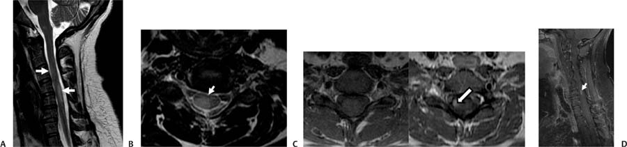
Case 86 A 42-year-old woman with paresthesias in both arms. (A) Sagittal T2-weighted image (WI) of the cord shows multiple oval areas of increased signal in the ventral and dorsal cord spanning the length of one to two vertebral bodies (arrows). (B) Axial T2WI of the cervical spine shows a wedge-shaped peripheral cord lesion dorsally (arrow). (C) Axial T1WI before and after gadolinium injection. Note the enhancement of one of the cord lesions (arrow). (D) Sagittal fat-suppressed T1WI shows a cigar-shaped enhancing cord lesion that spans less than the height of one vertebral body (arrow). • Multiple sclerosis (MS) in the cord: This is characterized by peripherally located focal cord lesions that are less than two vertebral segments in length and occupy less than half of the cross-sectional area of the cord. In the axial images, the lesions may have a wedge shape, with the base at the cord surface, or a round shape if there is no contact with the cord surface. • Devic neuromyelitis optica:
Clinical Presentation
Imaging Findings
Differential Diagnosis
![]()
Stay updated, free articles. Join our Telegram channel

Full access? Get Clinical Tree


