 Clinical Presentation
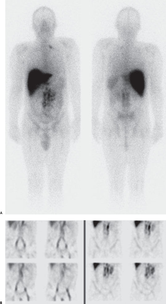
A 54-year-old man presents for cancer restaging.

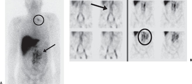
(A) Anterior and posterior whole-body imaging demonstrates intense physiologic uptake in the liver diffusely as well as milder blood pool, marrow, and bowel activity. Multifocal abnormal radiopharmaceutical uptake in the mid abdominal region (arrow), as well as focal uptake in the left supraclavicular region (circle), is seen. (B) Dual coronal SPECT imaging with Tc99m-RBCs (left images) at the same time as acquisition from the original tracer in A (right images) demonstrates abnormal accumulation in retroperitoneal aortocaval lymph nodes (circle), separate from the physiologic blood pool distribution mapped simultaneously on RBC scan (arrow).
 Differential Diagnosis
• ProstaScint scan with metastasis within the mid abdomen and left supraclavicular region:
Related posts:


Stay updated, free articles. Join our Telegram channel

Full access? Get Clinical Tree


