 Clinical Presentation
A 45-year-old man with fever and hemoptysis.
Further Work-up

 Imaging Findings

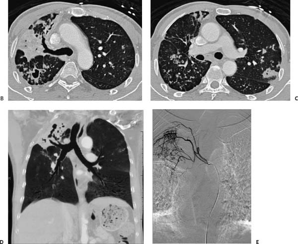
(A) Posteroanterior chest radiograph demonstrates dense right upper lobe consolidation with a suggestion of cavitation (arrows). (B) Contrast-enhanced computed tomography (CT) of the chest (lung windows) at the level of the aortic arch confirms the right upper lobe consolidation (arrow). There is a thickwalled, cavitary mass in the superior segment of the right lower lobe. It is surrounded by multiple centrilobular nodules and tree-in-bud opacity. (C) Contrastenhanced CT of the chest (lung windows) at the level of the pulmonary artery shows multiple nodules on the left, the largest of which appears cavitary (black arrow). On the right, there are diffusely scattered centrilobular nodules and areas of bronchial wall thickening (white arrows). (D) Minimum-intensity-projection CT of the chest (coronal re-formation) shows the right upper lobe consolidation and cavitation to advantage (arrow). (E
Stay updated, free articles. Join our Telegram channel

Full access? Get Clinical Tree


