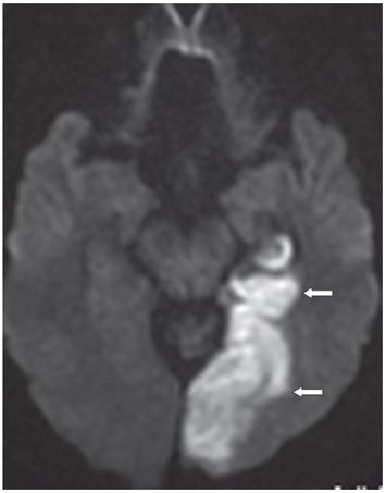
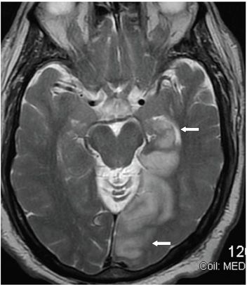
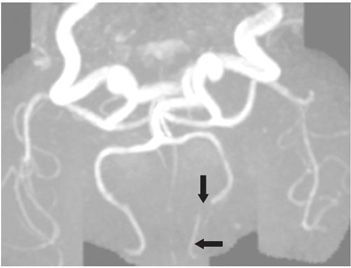
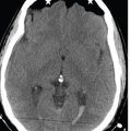
FINDINGS Figure 86-1. Axial NCCT through the occipital lobes. There is a large left occipital hypodensity (arrows) abutting the tentorium medially. Figure 86-2. Axial DWI through the occipital lobes. There is a large hyperintensity (low ADC not shown) consistent with area of restricted diffusion in the left occipital lobe. Figure 86-3. Axial T2WI through the occipital lobes. There is a large left occipital lobe hyperintensity extending into the posteromedial left temporal lobe (arrows). Figure 86-4. 3D TOF MRA. There is a loss of signal in the left posterior cerebral artery (PCA) at the P2–P3 junction (vertical arrow). Distally the left PCA is severely attenuated (transverse arrow).


Stay updated, free articles. Join our Telegram channel

Full access? Get Clinical Tree


