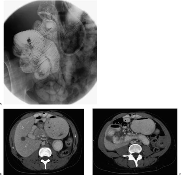
Case 86 An 18-year-old woman presents to the gastroenterology clinic with chronic, intermittent abdominal pain. (A) Small-bowel series shows multiple jejunal loops (arrows) within the right hemiabdomen, indicating malrotation. (B) Contrast-enhanced computed tomography (CT) shows a dilated loop of small bowel in the left hemiabdomen with mural thickening (arrow) and multiple small-bowel loops in the anterior right hemiabdomen (arrowhead). (C) The thickened loop leads into an anomalous jejunal course (arrows), crossing the midline adjacent to the duodenum and behind the superior mesenteric artery (fossa of Waldeyer) and continuing as multiple loops in the right hemiabdomen. • Paraduodenal (right) hernia: Paraduodenal (right) hernia with resulting malrotation is the most likely choice of diagnosis, given the thickened small-bowel loop and the abnormally positioned small bowel. • Malrotation: This is a possibility for the fluoroscopic study, but the CT findings and symptoms suggest herniation with ischemic changes.

 Clinical Presentation
 Imaging Findings

 Differential Diagnosis
Stay updated, free articles. Join our Telegram channel

Full access? Get Clinical Tree


