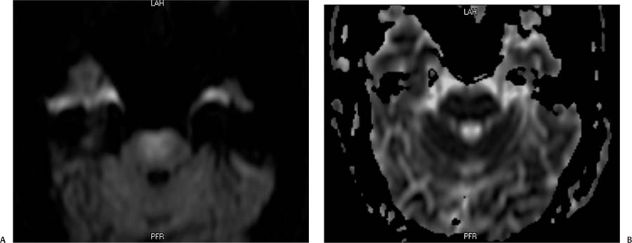
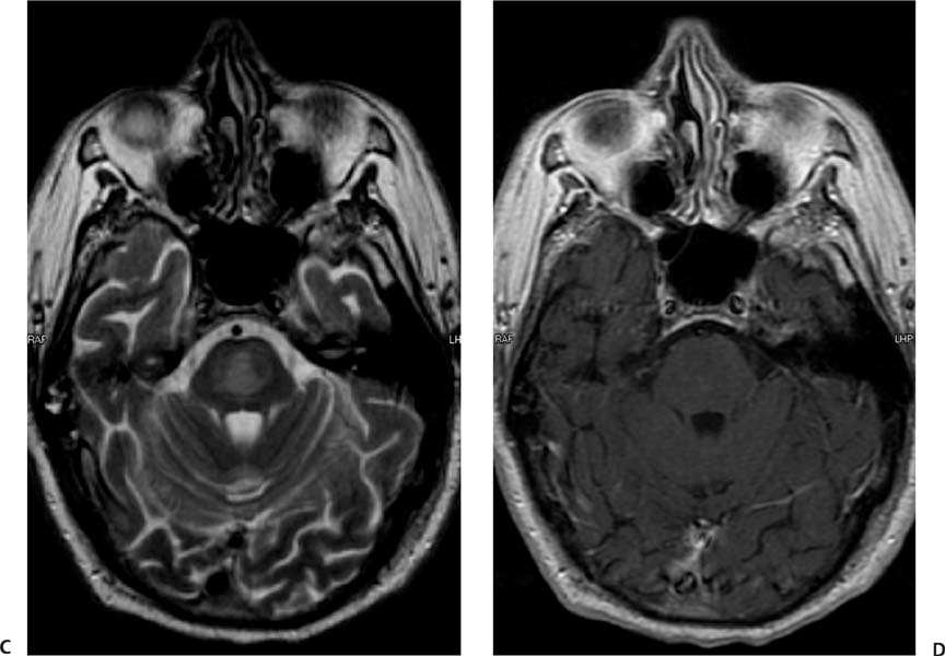
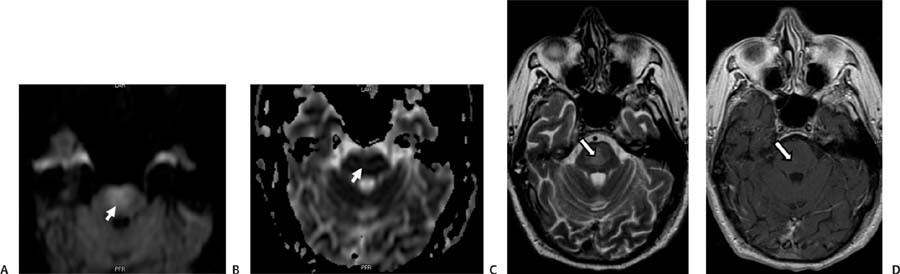
Case 87 A 68-year-old with the recent onset of spastic quadriparesis. (A) Diffusion-weighted image (WI) shows an area of increased signal without corresponding low signal on the apparent diffusion coefficient (ADC) map, consistent with T2 shine-through (arrow). (B) ADC map does not show decreased signal in the area of diffusion abnormality, indicating T2 shine-through (arrow). (C) Axial T2WI demonstrates an area of high signal in the central portion of the pons (arrow). (D) Axial T1WI after gadolinium contrast fails to demonstrate enhancement or T1 signal change. • Central pontine myelinolysis (CPM):
Clinical Presentation
Further Work-up
Imaging Findings
Differential Diagnosis
![]()
Stay updated, free articles. Join our Telegram channel

Full access? Get Clinical Tree


