 Clinical Presentation
A 37-year-old woman with chronic cough and infertility.
Further Work-up
 Imaging Findings
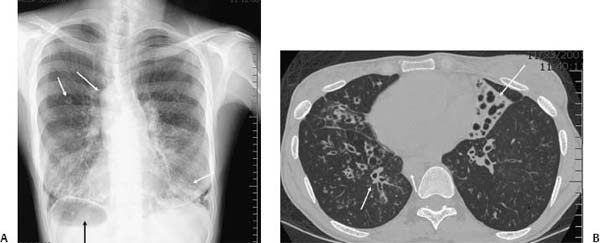
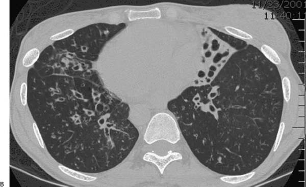
(A) Posteroanterior chest radiograph demonstrates bilateral lower lobe–predominant bronchiectasis and bronchial wall thickening. There is a basally predominant nodular interstitial pattern. The aortic arch, cardiac apex, and stomach bubble are right-sided (arrows). (B) Noncontrast computed tomography (CT) of the chest (lung windows) through the lung bases confirms widespread bronchiectasis and bronchial wall thickening. There are multiple centrilobular nodules and areas of tree-in-bud opacity. There is consolidation in the “left middle lobe” in this patient with situs inversus (arrows).
 Differential Diagnosis
• Primary ciliary dyskinesia:
Stay updated, free articles. Join our Telegram channel

Full access? Get Clinical Tree


