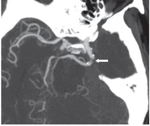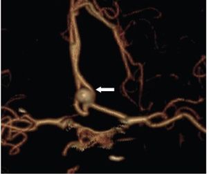

FINDINGS Figure 87-1. Oblique 3D volume-rendering CTA of the left ICA. There is a large vessel (vertical arrow) joining the left ICA to the mid-basilar artery. The surface nodularity is due to calcifications. The proximal basilar artery is not visualized (congenitally hypoplastic) while the distal basilar artery is robust (transverse arrow). Figure 87-2. Axial oblique MIP of the same vessel (arrow). There are multiple calcific foci on it. Figure 87-3. 3D volume rendering of the anterior circulation in the same patient. There is an anterior communicating artery aneurysm (arrow).
DIFFERENTIAL DIAGNOSIS N/A.
DIAGNOSIS Persistent trigeminal artery (PTA).
DISCUSSION
Stay updated, free articles. Join our Telegram channel

Full access? Get Clinical Tree


