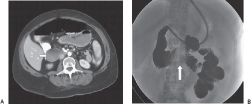
Case 87 A 25-year-old patient presents with midepigastric pain, vomiting, and weight loss. (A) Infused computed tomographic (CT) scan shows dilatation of the second portion of the duodenum (arrow) with constriction of the third portion between the superior mesenteric artery (SMA) and the aorta. (B) Upper gastrointestinal (GI) barium study shows dilatation of the fi rst and second portion of the duodenum with vertical bandlike constriction (arrow) of the third portion over the spine.
 Clinical Presentation
 Imaging Findings
 Differential Diagnosis
Stay updated, free articles. Join our Telegram channel

Full access? Get Clinical Tree


