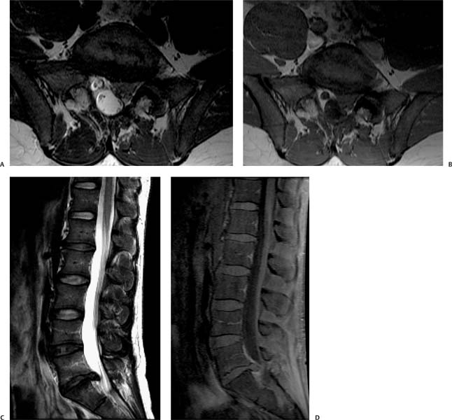
Case 88 A 52-year-old with numbness in the left foot. (A) Axial T2-weighted image (WI) at the level of the L5-S1 intervertebral disk shows an oval mass continuous with the disk occupying the left lateral recess (asterisk). Note the posterior displacement of the left S1 nerve root (arrowhead). (B) Axial T1WI at the level of the L5-S1 intervertebral disk. Note the thin plane of fat that separates the lesion from the facet joint (arrow). (C) Observe the continuity of the lesion with the L5-S1 disk on the sagittal T2WI (arrow). (D) Sagittal fat-suppressed T1WI shows enhancement of the epidural plexus (arrow), but not of the disk herniation. • Disk herniation: This is a localized displacement of disk material beyond the limits of the intervertebral disk space. It encroaches on the epidural fat and sometimes the dural sac and nerve roots. The herniation is usually contiguous with the rest of the disk, but free fragments are possible. The herniation is usually isointense to the parent disk on T1- and T2-weighted sequences.
Clinical Presentation
Imaging Findings
Differential Diagnosis
Stay updated, free articles. Join our Telegram channel

Full access? Get Clinical Tree


