 Clinical Presentation
A 21-year-old man with chronic cough.
Further Work-up
 Imaging Findings
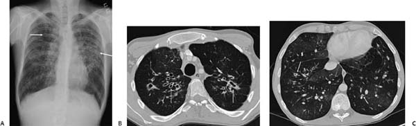
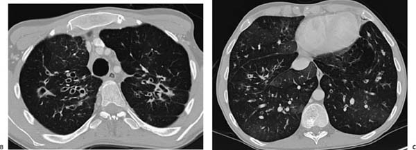
(A) Posteroanterior chest radiograph demonstrates bilateral upper lobe–predominant bronchial wall thickening and bronchiectasis (arrows). The lung volumes are relatively large. (B) Contrast-enhanced computed tomography (CT) through the upper lobes confirms upper lobe bronchial wall thickening and bronchiectasis (arrows). (C
Stay updated, free articles. Join our Telegram channel

Full access? Get Clinical Tree


