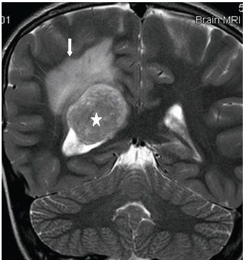
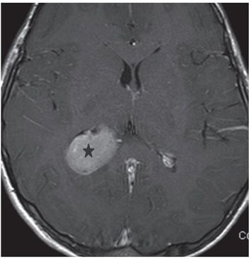
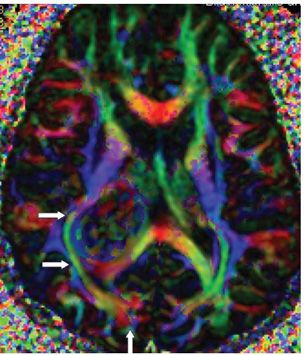
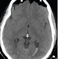
FINDINGS Figure 88-1. Axial FLAIR through the trigones. There is a homogeneous isointense (to gray matter [GM]) mass within the right trigone measuring 3.8 cm × 2.6 cm at the widest point (star). There is periventricular hyperintensity extending into the white matter (WM) consistent with parenchymal vasogenic edema (arrow). Figure 88-2. Coronal T2WI through the mass. Mass (star) is mildly heterogeneous but isointense to GM. Peripherally superiorly and medially the mass is as hyperintense as the adjacent periventricular hyperintense vasogenic edema (arrow). Figure 88-3. Post-contrast T1WI. There is homogeneous contrast enhancement of the smooth marginated mass (star). Figure 88-4. DTI color directional map. There is mass effect on the right inferior fronto-occipital fasciculus and inferior longitudinal fasciculus (transverse arrows) which have been compressed laterally. There is also posterior displacement of the fibers in the right splenium with disorganization of the right forceps major (vertical arrow).
DIFFERENTIAL DIAGNOSIS Choroid plexus carcinoma (CPC), choroid plexus papilloma (CPP), intraventricular meningioma, ependymoma.
DIAGNOSIS


Stay updated, free articles. Join our Telegram channel

Full access? Get Clinical Tree


