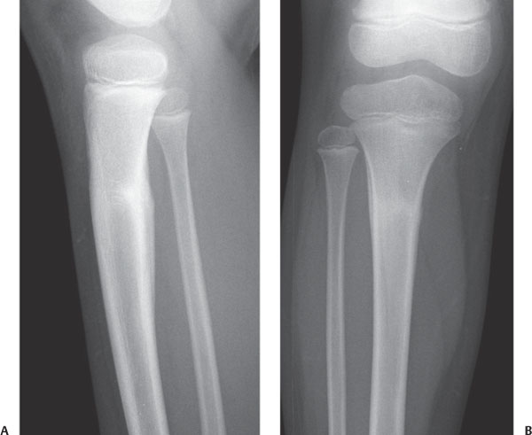
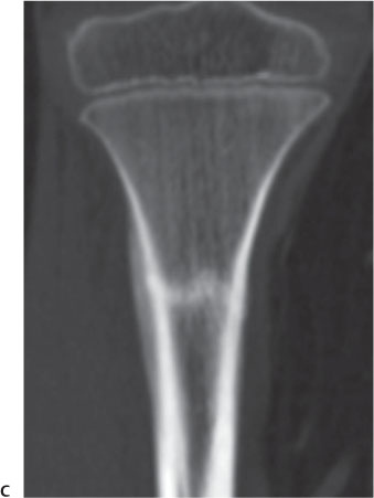
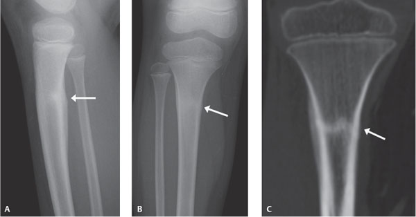
Case 88 Persistent calf pain in an athlete. (A,B) Frontal and lateral plain radiographs of the proximal tibia and fibula demonstrate a transverse band of sclerosis extending across the proximal tibia, with an associated periosteal reaction (arrows). (C) Coronal reformat computed tomography image of the proximal tibia again demonstrates the transverse sclerosis and periosteal reaction of the proximal tibia (arrow).
Clinical Presentation
Further Work-up
Imaging Findings
Differential Diagnosis
Stay updated, free articles. Join our Telegram channel

Full access? Get Clinical Tree


