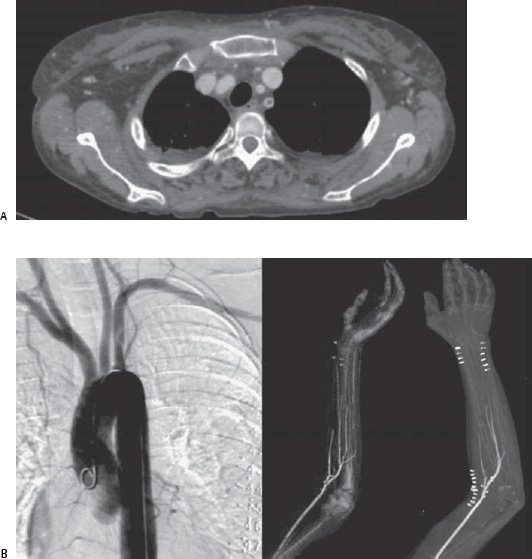
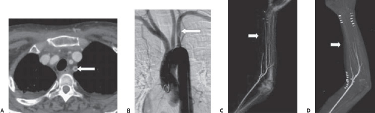
Case 88 A 35-year-old man presents to the emergency department with an abrupt onset of pain and pallor in the left arm. (A) Infused computed tomographic (CT) scan shows filling defect within the proximal portion of the left subclavianartery (arrow). (B) Angiogram of the aortic arch shows subclavian artery filling defect proximal to the takeoff of the ver tebral artery (arrow). (C,D) Three-dimensional volume-rendered and maximal-intensity-projection images from a computed tomographic angiogram (CTA) of the left arm show abrupt occlusion of the forearm trifurcation arteries, indicating distal thromboembolism (arrows). Surgical clips indicate attempted surgical revascularization.
 Clinical Presentation
 Imaging Findings
 Differential Diagnosis
Stay updated, free articles. Join our Telegram channel

Full access? Get Clinical Tree


