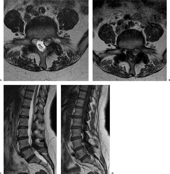
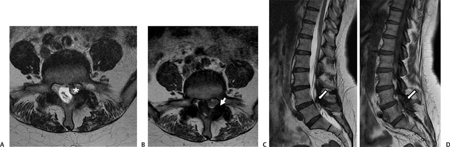
Case 89 A 46-year-old with weakness and pain in the left leg. (A) Axial T1-weighted image (WI) at the level of L4-L5 demonstrates an oval lesion in the left lateral recess with a rim of low signal and central heterogeneous high signal (asterisk). The left L5 nerve root cannot be separated from the mass. (B) Axial T1WI shows heterogeneous high signal in the left lateral recess lesion, which abuts the facet joint (arrow). (C) Sagittal T2WI shows the close relationship of the lesion with the facet joint (arrow). There is no contact with the intervertebral disk. (D) Sagittal T1WI shows the close relationship of the lesion with the facet joint (arrow). There is no contact with the intervertebral disk. • Facet synovial cyst:
Clinical Presentation
Imaging Findings
Differential Diagnosis
![]()
Stay updated, free articles. Join our Telegram channel

Full access? Get Clinical Tree


