Case 89
Case History
A 49-year-old woman presents with left breast calcifications that are new since her previous screening mammogram 4 years ago.
Physical Examination
• normal exam
Mammogram
Calcifications (Figs. 89–1 and 89–2)
• type: amorphous/indistinct
• distribution: segmental
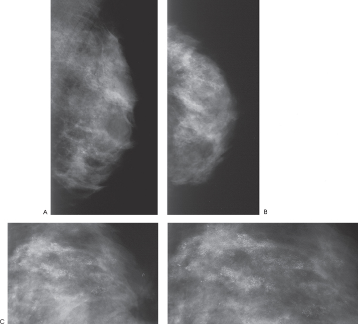
Figure 89–1. Amorphous calcifications are present throughout the entire left upper outer quadrant. (A). Left MLO mammogram. (B). Left CC mammogram. (C). Left LM magnification mammogram.