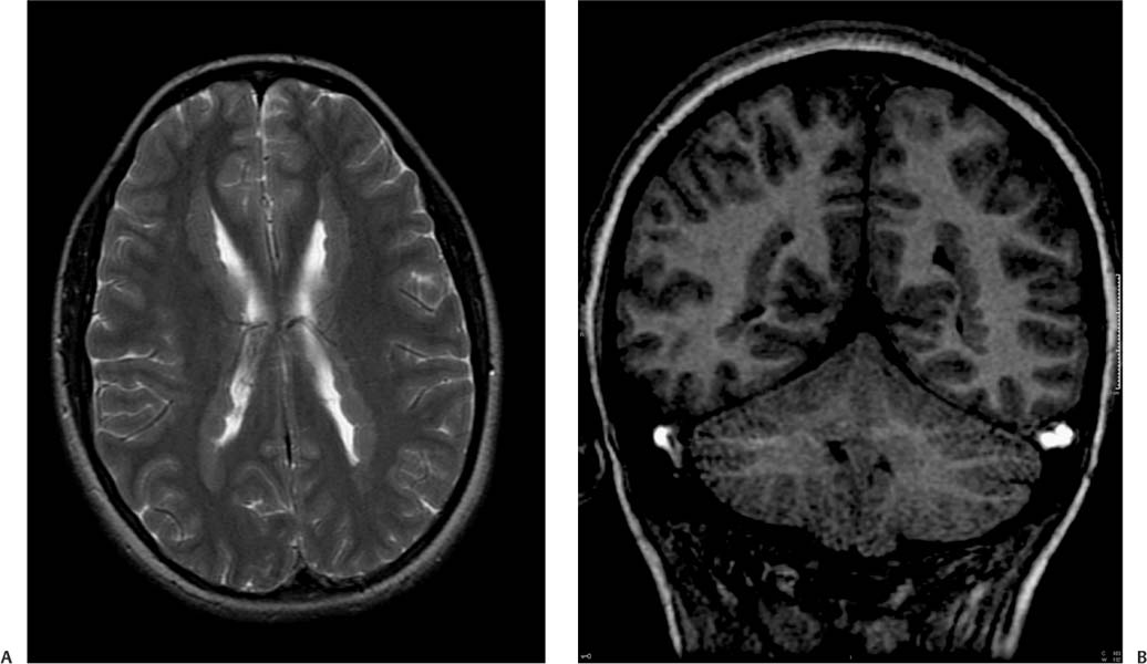
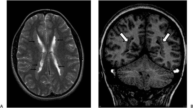
Case 9 A patient undergoing a work-up for epilepsy. (A) Axial T2-weighted image (WI) of the brain demonstrates multiple nodules, with signal intensity similar to that of the cerebral cortex, lining the lateral ventricles (arrows). There is a serrated appearance of the inner margin of the ventricles. (B) Coronal T1WI confirms the presence of nodules along the ventricular walls. The nodules have signal intensity like that of the cortex (arrows). • Subependymal heterotopia: Nodules of gray matter along the ventricular surface are suggestive of subependymal heterotopia. This may appear exophytic, extending to the ventricle. • Subependymal nodules of tuberous sclerosis: These are irregularly shaped and often calcified. They are not isointense to cortex. They may enhance. • Band heterotopia:
Clinical Presentation
Imaging Findings
Differential Diagnosis
![]()
Stay updated, free articles. Join our Telegram channel

Full access? Get Clinical Tree


