 Clinical Presentation
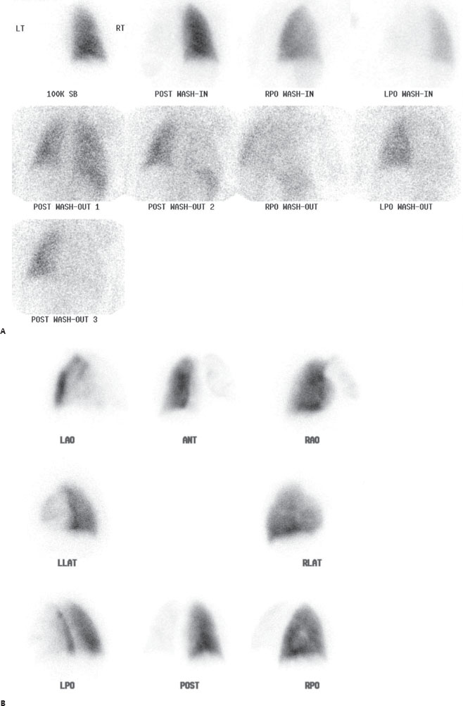
A 59-year-old woman with shortness of breath. Chest radiographs were unremarkable.

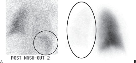
(A) Xenon 133 ventilation scintigraphy demonstrates decreased ventilation to the entire left lung on single breath and wash-in. There is eventual equilibration but then abnormal retention throughout the entire left lung on washout. More focal retention of radiotracer is seen inferiorly on the right (circle). (B) Tc99m MAA perfusion images demonstrate markedly decreased perfusion to the entire left lung (circle).
 Differential Diagnosis
• Low probability for pulmonary embolism (PE) and xenon retention in a fatty liver: Matched perfusion defects, even if large, with normal chest x-ray (CXR) findings are always low (or very low) probability. The right lower Xe133 activity is below the diaphragm and the classic appearance for a fatty liver.
• Ventilatory retention at the right lung base:


Stay updated, free articles. Join our Telegram channel

Full access? Get Clinical Tree


