 Clinical Presentation
A 58-year-old man with progressive dyspnea, cough, and dysphagia.
 Imaging Findings
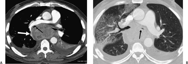
(A) Contrast-enhanced computed tomography (CT) shows a large mediastinal mass (white arrow) with heterogeneous enhancement, airway compression (black arrows), and bilateral pleural fluid collections. (B) Lung window image shows interlobular septal thickening and ground-glass opacity in the right lung.
 Differential Diagnosis
• Small-cell lung cancer (SCLC): The most common imaging presentation is a large mediastinal mass, generally without imaging evidence of a lung parenchymal lesion.
• Lymphoma: A large mediastinal mass may also be the initial presentation of a lymphoma. The presence of airway obstruction helps in the differentiation between lymphoma and SCLC. Bronchial narrowing or an intraluminal mass is more common in lung cancer than in lymphoma.
Stay updated, free articles. Join our Telegram channel

Full access? Get Clinical Tree


