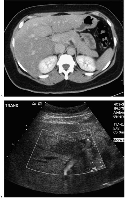
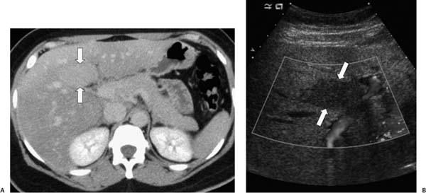
Case 9 A 54-year-old man presents with vague abdominal symptoms and weight loss. (A) Infused abdominal computed tomography (CT) shows hypoattenuation of the liver sparing an ovoid portion (arrows) of the medial segment of the left lobe. (B) Ultrasound shows this ovoid region (arrows) to be adjacent to the portal vein, well defined, and hypoechoic compared with the remainder of the liver. • Focal fatty sparing: This is the most likely diagnosis for a well-defined, relatively hyperdense region of the medial segment of the left lobe in an otherwise hypodense liver suspicious for hepatic steatosis. The periportal location and absence of mass effect further support this diagnosis. • Hepatic tumor:
 Clinical Presentation
 Imaging Findings
 Differential Diagnosis
![]()
Stay updated, free articles. Join our Telegram channel

Full access? Get Clinical Tree


