 Clinical Presentation
A 50-year-old woman with severe dyspnea and an elevated angiotensin-converting enzyme level.
Further Work-up
 Imaging Findings
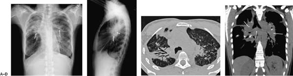
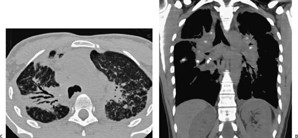
(A) Posteroanterior chest radiograph demonstrates hilar and paratracheal lymphadenopathy with upper lobe retraction (arrows). (B) Lateral chest radiograph shows the lymphadenopathy to advantage (arrows). (C) Computed tomography (CT) of the chest (lung windows) shows areas of consolidation with traction bronchiectasis on the right (right arrows). In the left upper lobe, there are widespread perilymphatic nodules and thickening of the bronchovascular bundles (left arrow). (D) CT of the chest (coronal, soft-tissue window) best shows the extensive calcified lymphadenopathy (arrows).
 Differential Diagnosis
• Sarcoidosis: Calcified hilar and mediastinal lymphadenopathy and upper lobe–predominant perilymphatic nodules are suggestive of sarcoidosis.
• Idiopathic pulmonary fibrosis:
Stay updated, free articles. Join our Telegram channel

Full access? Get Clinical Tree


