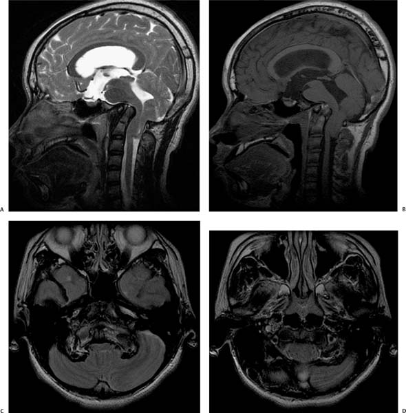
Case 91 A 17-year-old with a history of hydrocephalus. (A) Sagittal T2-weighted image (WI) of the brain: The basal angle measures 176 degrees. This angle is subtended by the junction of the nasion-tuberculum and tuberculum-basion tangents. Average is 134 to 135 degrees, minimum is 121 degrees, and maximum is 148 to 149 degrees. Consider platybasia if the angle is greater than 150 degrees. (B) Sagittal T1WI of the brain: The Chamberlain line extends from the posterior margin of the hard palate to the opisthion (posterior margin of the foramen magnum). It is abnormal if the tip of the odontoid projects more than 5 mm above the Chamberlain line. (C) Axial T2WI at the level of the foramen magnum demonstrates the tip of the dens (asterisk) at the same level as the occipital condyles (arrows). Note the compressive effect on the medulla oblongata. (D) Axial T2WI again demonstrates the compressive effect on the medulla oblongata and on the cerebellar tonsils (arrowheads), which are low-lying. • Basilar invagination and platybasia:
Clinical Presentation
Imaging Findings
Differential Diagnosis
![]()
Stay updated, free articles. Join our Telegram channel

Full access? Get Clinical Tree


