 Clinical Presentation
An 80-year-old man with cough and dyspnea.
Further Work-up
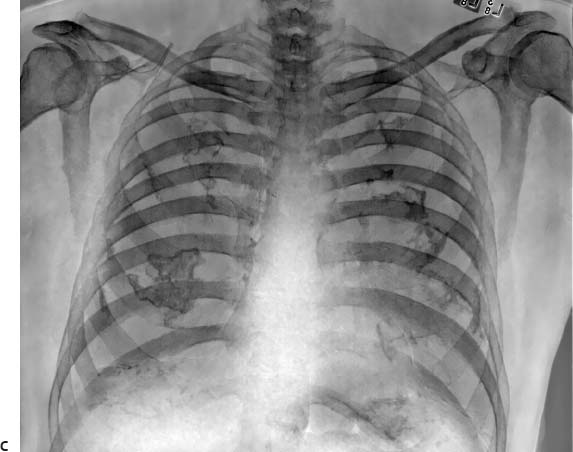
 Imaging Findings
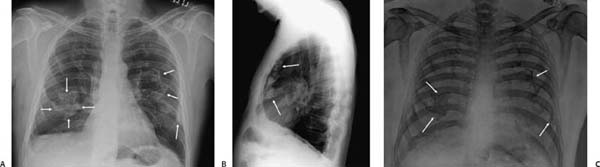
(A) Posteroanterior (PA) chest radiograph demonstrates irregularly shaped areas of calcification bilaterally (arrows). The diaphragmatic surfaces are also involved. (B) Lateral chest radiograph shows that the calcification is distributed along the pleural surfaces (arrows). (C) Energy-subtracted views from the PA radiograph show the calcification to advantage (arrows).
 Differential Diagnosis
Stay updated, free articles. Join our Telegram channel

Full access? Get Clinical Tree


