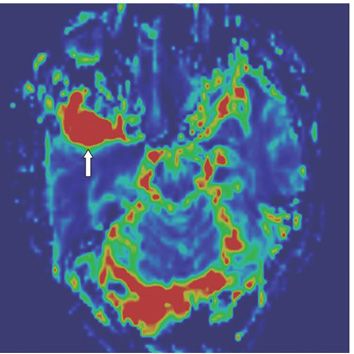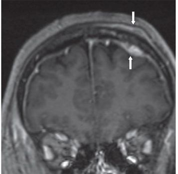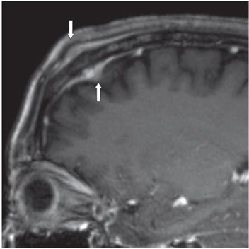


FINDINGS Figure 91-1. Axial post-contrast T1WI through the temporal lobes. There is a homogenously enhancing extraaxial mass within the right middle cranial fossa (arrow). Figure 91-2. Cerebral blood volume (CBV) map from dynamic susceptibility contrast MR perfusion. There is markedly elevated relative Cerebral Blood Volume (rCBV) within the right middle cranial fossa mass (arrow). Figures 91-3 and 91-4. Coronal and sagittal reformatted post-contrast 3D T1WI through the frontal lobes, respectively. There is a concurrent dural-based enhancing mass with dural tails at the left frontal convexity with contiguous osseous and extracranial components (arrows).
DIFFERENTIAL DIAGNOSIS Meningioma, lymphoma, hemangiopericytoma, metastases.
DIAGNOSIS
Stay updated, free articles. Join our Telegram channel

Full access? Get Clinical Tree


