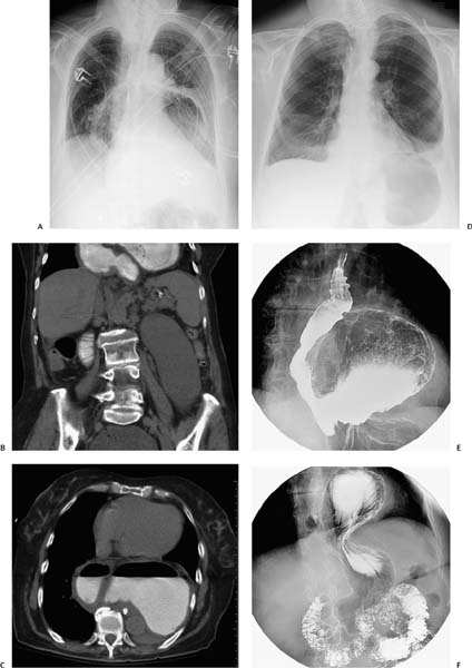
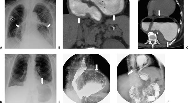
Case 91 For the following two patients with midepigastric pain, what is the differential diagnosis for the radiographic findings? What are the diagnosis and clinical significance in each case? (A) Radiograph of the first patient shows an air-filled structure (arrows) in the chest. (B) Coronal computed tomography (CT) with oral contrast shows an intrathoracic stomach (arrows). Barium within the stomach indicates absence of complete volvulus, although partial organoaxial volvulus is present (see below). (C) Axial CT shows a nasogastric (NG) tube passing posterior to the stomach (arrows) toward the gastroesophageal junction (GEJ). (D) Radiograph of the second patient shows a large, air-filled structure in the left upper quadrant/chest (arrow). (E) Barium study shows near-normal position of the GEJ and a large paraesophageal hernia (arrow). (F)
 Clinical Presentation
 Imaging Findings
![]()
Stay updated, free articles. Join our Telegram channel

Full access? Get Clinical Tree


