CASE 91
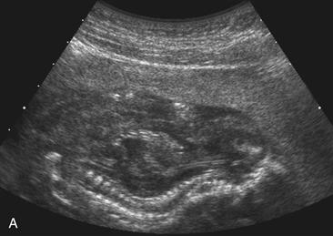
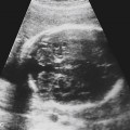
Used with permission from McGahan JP, Benacerraf BR: Fetal abdomen and pelvis. In McGahan JP, Goldberg BB [eds]: Diagnostic Ultrasound, 2nd ed. New York: Informa Healthcare USA, 2008.
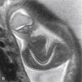
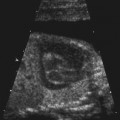
Used with permission from Anderson Publishing Ltd. from Victoria T, et al: Fetal MRI of common non-CNS abnormalities: A review. Appl Radiol 2011;40[6]:8-17. © Anderson Publishing Ltd.
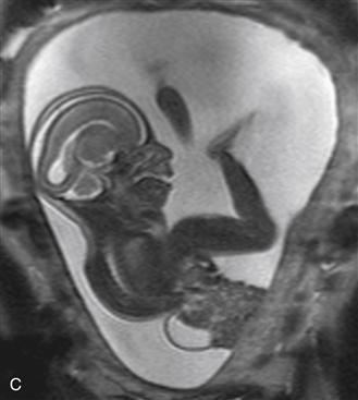
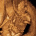
Used with permission from Anderson Publishing Ltd. from Victoria T, et al: Fetal MRI of common non-CNS abnormalities: A review. Appl Radiol 2011;40[6]:8-17. © Anderson Publishing Ltd.
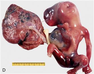
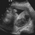
Used with permission from McGahan JP, Benacerraf BR: Fetal abdomen and pelvis. In McGahan JP, Goldberg BB [eds]: Diagnostic Ultrasound, 2nd ed. New York: Informa Healthcare USA, 2008.
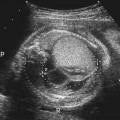
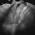
History: A patient undergoes a prenatal ultrasound scan that shows an unusual appearance to the fetus.
1. What should be included in the differential diagnosis for Figure A? (Choose all that apply.)
D. Trisomy 21
2. What is the most common genetic abnormality that occurs with limb–body wall complex?
B. Trisomy 18
C. Trisomy 21
D. XO Turner syndrome karyotype
Stay updated, free articles. Join our Telegram channel

Full access? Get Clinical Tree


