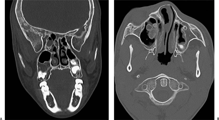
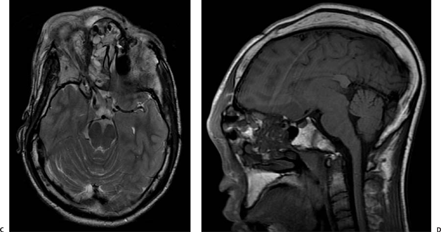
Case 92 A 36-year-old woman with long-standing cosmetic deformity of the face. (A) Reformatted coronal computed tomography (CT) of the face shows replacement and enlargement of the bone marrow in the greater sphenoid wing and maxillary bones (asterisks), predominantly on the right, with a ground glass attenuation. Note the narrowing of the right superior orbital fissure (arrow) in comparison with the left. (B) Axial CT shows proliferation of abnormal bone marrow within the right maxillary sinus and lateral wall of the left maxillary sinus (arrows). Asymmetry of the face is evident. (C) Axial T2-weighted image (WI) through the orbital roof shows heterogeneous signal of the abnormal bone marrow. Note the elongation of the right optic nerve (arrows) and proptosis, caused by the enlarged orbital walls. (D) Sagittal T1WI shows that the areas of marrow replacement display relatively low signal (arrow). Areas of high T1 signal within the ethmoid complex may represent secretions or secondary retention of mucus (arrowhead). Note the magnetic susceptibility artifact in the region of the nasion, secondary to prior surgery.
Clinical Presentation
Further Work-up
Imaging Findings
Stay updated, free articles. Join our Telegram channel

Full access? Get Clinical Tree


