 Clinical Presentation
An 80-year-old woman with dyspnea and a positive purified protein derivative test result.
Further Work-up
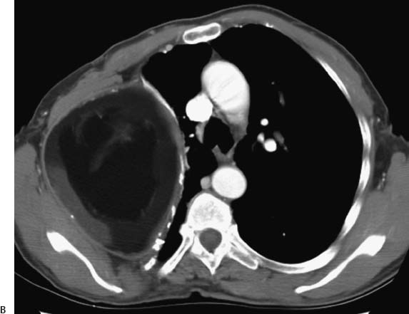
 Imaging Findings
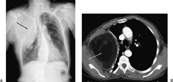
(A) Chest radiograph demonstrates severe deformity of the right chest and ribs, multiple surgical clips, right-sided volume loss, and a large mass in the right uppes hemithorax (arrow). (B) Contrast-enhanced computed tomography (soft-tissue windows) shows a heterogeneous but predominant fat density mass in the right hemithorax (arrow).
Stay updated, free articles. Join our Telegram channel

Full access? Get Clinical Tree


