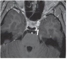
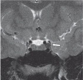
FINDINGS Figure 92-1. Coronal post-contrast T1WI shows an enhancing small mass (arrow) in the expected course of the left oculomotor nerve in the superolateral left cavernous sinus. Figure 92-2. Axial post-contrast T1WI shows the anterior location of the mass (arrow) within the left cavernous sinus. Figure 92-3. Coronal T2WI through the cavernous sinuses in a companion case. There is a well-defined round hypointense mass (arrow) in the mid-left cavernous sinus.
DIFFERENTIAL DIAGNOSIS Meningioma, metastasis (including perineural spread), lymphoma, schwannoma, inflammatory pseudotumor.
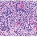
DIAGNOSIS Cavernous sinus schwannoma (oculomotor nerve).
DISCUSSION


Stay updated, free articles. Join our Telegram channel

Full access? Get Clinical Tree


