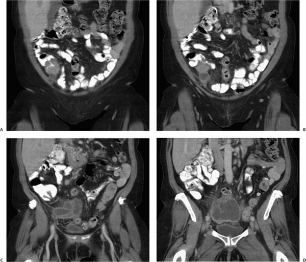
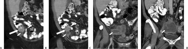
Case 92 A 75-year-old woman presents with pelvic pain and is found to have fever and leukocytosis. (A) Coronal reformatted infused computed tomography shows a mass (large arrow) in the cecum with adjacent mural thickening. A fluid-density structure (small arrow) abutting this mass may represent the base of a dilated appendix. The barium-filled terminal ileum is annotated (arrowhead). (B) More posterior image shows the mass (large arrow), the base of the appendix (small arrow), the ileocecal valve (large arrowhead), and pericecal fat stranding (small arrowhead). (C) More posterior image shows the dilated appendix (arrow). (D)
 Clinical Presentation
 Imaging Findings
![]()
Stay updated, free articles. Join our Telegram channel

Full access? Get Clinical Tree


