CASE 92
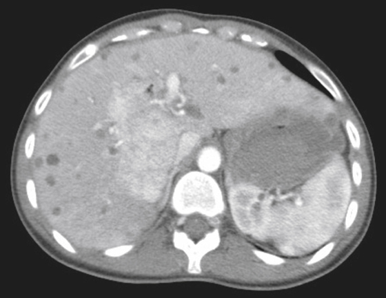
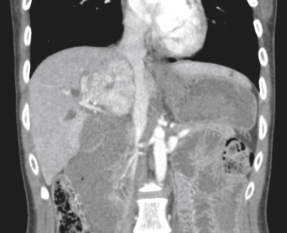
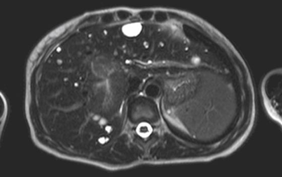
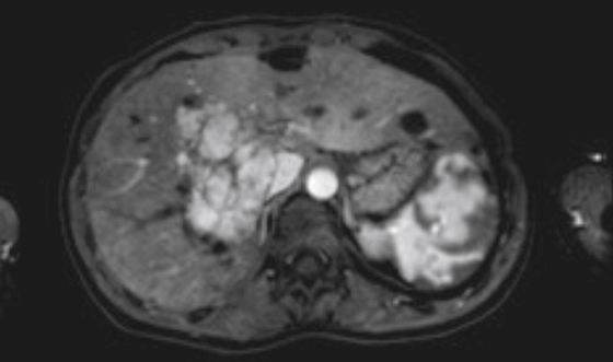
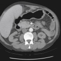
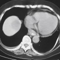
History: A 32-year-old woman presents with chest pain.
1. Which of the following should be included in the differential diagnosis of the imaging finding shown in the figures? (Choose all that apply.)
2. Why do focal nodular hyperplasia (FNH) lesions take up sulfur colloid?
A. The lesion contains hepatocytes.
B. The lesion contains Kupffer cells.
C. The lesion contains blood pools.
D. The lesion is hypervascular.
3. In what demographic group is FNH most commonly seen?
Stay updated, free articles. Join our Telegram channel

Full access? Get Clinical Tree


