Case 92
Case History
A 64-year-old woman with new right breast calcifications.
Physical Examination
• Normal exam
Mammogram
Calcifications (Figs. 92–1 and 92–2)
• type: amorphous/indistinct
• distribution: grouped/clustered
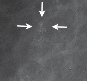
Figure 92–1. Right MLO magnification mammogram: Faint amorphous calcifications (arrows) are present in the upper breast.
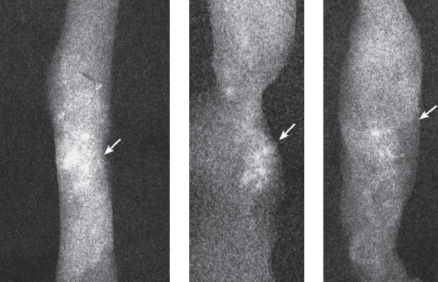
Figure 92–2. Specimen radiograph of percutaneous core right breast biopsy. Cores from the biopsy of the calcifications identified in Figure 92–1


Stay updated, free articles. Join our Telegram channel

Full access? Get Clinical Tree


