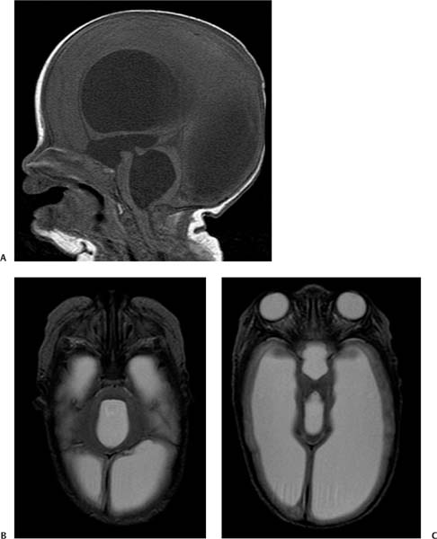
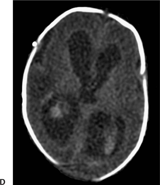
Case 93 A 30-week premature infant, now 3 months of age, presenting with increased head circumference. (A) Sagittal T1-weighted image (WI) shows dilatation of the entire ventricular system. Note the elongation and thinning of the corpus callosum (arrowheads), descent of the floor of the 3rd ventricle, which is obliterating the suprasellar cistern and abutting the pituitary gland (white arrow), and enlargement of the aqueduct (black arrow) and 4th ventricle. (B) Axial T2WI shows that the anteroposterior diameter of the 4th ventricle (asterisk) is significantly increased, resulting in flattening of the dorsal surface of the pons. (C) Axial T2WI shows that the dilated 3rd ventricle occupies the suprasellar cistern. The hypothalamic structures are splayed (arrows). (D) Computed tomography of the brain obtained 3 months earlier, when the patient was 2 weeks old. Intraventricular hemorrhage and mild ventricular dilatation are evident. Note the parenchymal hemorrhage in the right periventricular white matter (arrow). • Communicating hydrocephalus:
Clinical Presentation
Further Work-up
Imaging Findings
Differential Diagnosis
![]()
Stay updated, free articles. Join our Telegram channel

Full access? Get Clinical Tree


