 Clinical Presentation
A 52-year-old man with dyspnea on exertion.
Further Work-up

 Imaging Findings

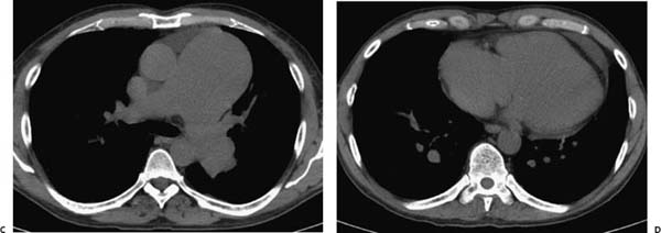
(A) Posteroanterior chest radiograph demonstrates cardiomegaly and massive central pulmonary artery enlargement (arrows). There is no evidence of pulmonary edema or pleural effusion. (B) Lateral chest radiograph confirms dilated right and left main pulmonary arteries (black arrows). Note the filling of the retrosternal clear space, a sign of right ventricular enlargement (white arrow). (C) Noncontrast computed tomography (CT) of the chest (soft-tissue windows) at the level of the main pulmonary artery confirms the significantly enlarged main pulmonary artery (arrows). Note the size discordance in comparison with the aorta. (D) Noncontrast CT of the chest (soft-tissue windows) through the heart shows the right-sided cardiac enlargement to advantage (arrows).
 Differential Diagnosis
• Pulmonary hypertension
Stay updated, free articles. Join our Telegram channel

Full access? Get Clinical Tree


