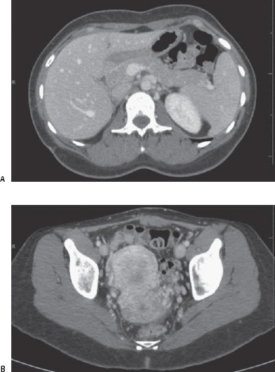
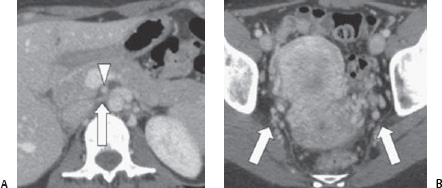
Case 93 A 37-year-old woman presents with chronic pelvic pain and hematuria. (A) Contrast-enhanced computed tomographic (CT) scan shows compression of the left renal vein (arrow) as it passes behind the superior mesenteric artery (SMA: arrowhead). (B) Pelvic image shows multiple dilated veins on both sides (arrows).
 Clinical Presentation
 Imaging Findings
 Differential Diagnosis
Stay updated, free articles. Join our Telegram channel

Full access? Get Clinical Tree


