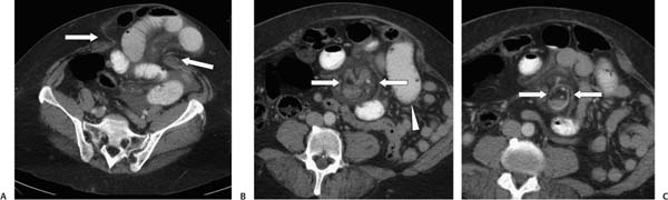
Case 93 A 62-year-old woman presents with abdominal pain and distension. (A) Contrast-enhanced computed tomography (CT) shows a wide-mouthed ventral hernia (arrows) containing both colon and small bowel. There is associated small-bowel dilatation, adjacent fat stranding, and a small amount of ascites. (B) Abdominal CT shows a rounded focus (arrows) containing small bowel, mesenteric fat, and mesenteric vessels. There is an adjacent, dilated small-bowel loop (arrowhead). (C)
 Clinical Presentation
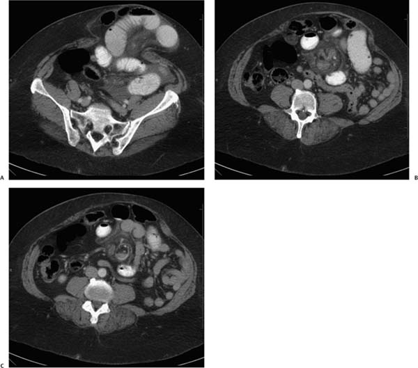
 Imaging Findings
![]()
Stay updated, free articles. Join our Telegram channel

Full access? Get Clinical Tree


