Case 93
Case History
A 43-year-old woman initially presented at an outside institution with a pea-sized palpable mass near the right nipple and calcifications on her mammogram. The mass was biopsied at the outside institution.
Physical Examination
• right breast: biopsy ecchymosis near right nipple; original nodule no longer detectable
• left breast: normal exam
Mammogram
Calcifications (Fig. 93–1)
• type: amorphous/indistinct
• distribution: grouped/clustered
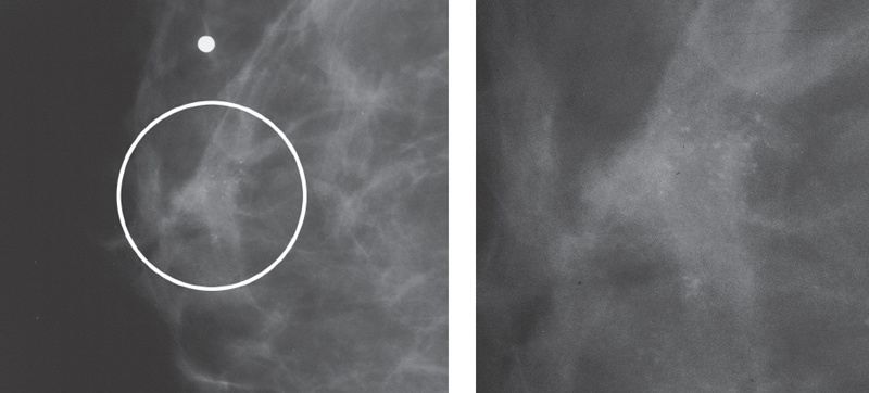
Figure 93–1. Right ML magnification mammogram: In the subareolar region there is a palpable lump marked by a radiopaque marker. The lump is associated with a cluster of amorphous calcifications (circled).
Ultrasound
Frequency
• 13 MHz
Mass


Stay updated, free articles. Join our Telegram channel

Full access? Get Clinical Tree


