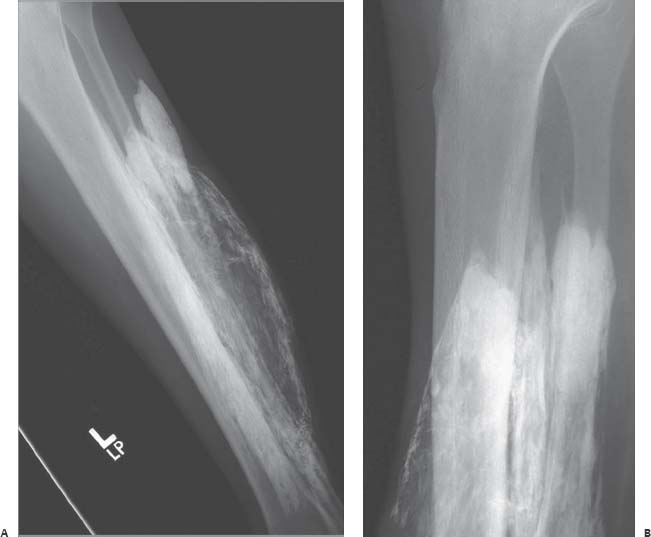
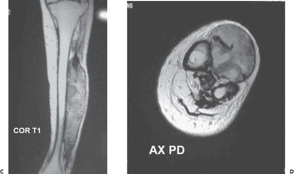
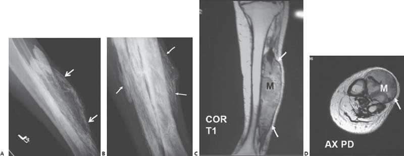
Case 94 A 60-year-old man presents with a several-year history of a slowly enlarging fluctuant mass in the left leg with a remote history of trauma. Further Work-up (A,B) Radiographs of the left tibia and fibula show a mineralized mass overlying the anterior compartment of the mid to distal leg. The calcifications are linear and platelike in morphology (arrows), with no bony matrix. (C,D) Magnetic resonance (MR) images show a fusiform, well-marginated soft-tissue mass (M) replacing the anterior compartment musculature. A peripheral rim of low signal intensity (arrows

 Clinical Presentation

 Imaging Findings

![]()
Stay updated, free articles. Join our Telegram channel

Full access? Get Clinical Tree


