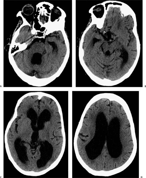
Case 94 A 72-year-old with gait disturbance and memory loss. (A) Axial computed tomography (CT) at the level of the pons shows dilatation of the 4th ventricle (asterisk). Note the prominence of the left temporal horn. (B) At the level of the midbrain, a large aqueduct (black arrow) and splaying of the hypothalamic structures (white arrows) are demonstrated. (C) There is fusiform dilatation of the 3rd ventricle (asterisk) and a round configuration of the frontal and occipital horns (white arrows). Note the small size of the sylvian fissures relative to the enlarged ventricles. (D)
Clinical Presentation
Imaging Findings
![]()
Stay updated, free articles. Join our Telegram channel

Full access? Get Clinical Tree


