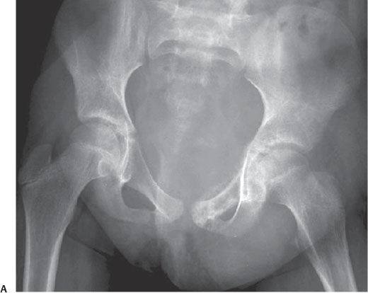
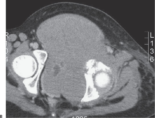
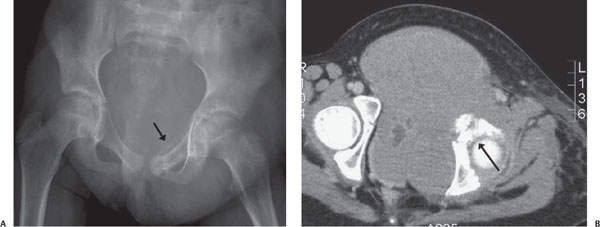
Case 94 A 9-year-old with progressive hip pain. (A) Anteroposterior radiograph of the pelvis: there is an aggressive lesion in the left superior pubic ramus with a periosteal reaction (arrow) and an associated soft-tissue mass. (B) Axial computed tomography (CT) image: there is destruction of the left acetabulum (arrow), with an associated large soft-tissue mass. • Ewing sarcoma: These findings are typical of Ewing sarcoma. • Osteosarcoma (OS): Typically presents as a large, mixed sclerotic/lytic lesion with a cloudlike matrix. It is less likely to occur in flat bones. • Chondrosarcoma: Most commonly occurs in patients older than 40 years and rarely in children.
Clinical Presentation
Further Work-up
Imaging Findings
Differential Diagnosis
Essential Facts
Stay updated, free articles. Join our Telegram channel

Full access? Get Clinical Tree


