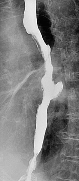
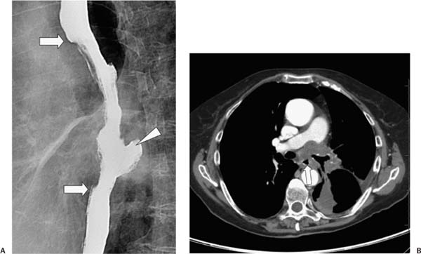
Case 94 A 69-year-old man has dysphagia. (A) Single-contrast barium study shows a long, irregularly marginated midesophageal stricture (arrows). A focal, posterolateral outpouching (arrowhead) of contrast is also visible. (B) Contrast-enhanced computed tomography (CT) shows a soft-tissue mass infiltrating the mediastinum at the level of the carina and narrowing the esophageal lumen (arrow).
 Clinical Presentation
 Imaging Findings
 Differential Diagnosis
Stay updated, free articles. Join our Telegram channel

Full access? Get Clinical Tree


