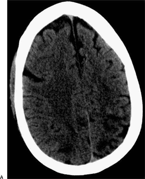
Case 95 A patient who has a history of lung cancer treated with chemotherapy now presents with headache and altered mentation. (A) Axial computed tomography (CT) of the brain shows areas of low attenuation in the subcortical white matter of both parietal lobes, without mass effect (arrows). (B) Axial fluid-attenuated inversion recovery (FLAIR) image demonstrates vasogenic edema in the frontal and parietal subcortical white matter bilaterally (arrows). (C) Coronal FLAIR image demonstrates vasogenic edema in the frontal and parietal subcortical white matter bilaterally (arrows). (D) Axial T1-weighted images (WIs) before and after gadolinium injection fail to demonstrate contrast enhancement (arrows). • Posterior reversible encephalopathy syndrome (PRES): Vasogenic edema is seen in the subcortical white mater of both parietal and occipital regions. There is no abnormal enhancement or significant mass effect.
Clinical Presentation
Further Work-up
Imaging Findings
Differential Diagnosis
Stay updated, free articles. Join our Telegram channel

Full access? Get Clinical Tree


