 Clinical Presentation
A 22-year-old woman with decreased exercise tolerance.
Further Work-up

 Imaging Findings

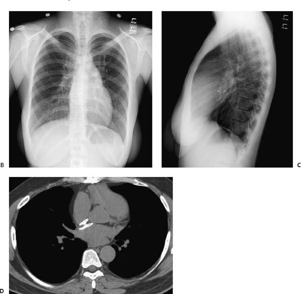
(A) Posteroanterior (PA) chest radiograph demonstrates enlargement of the main and central pulmonary arteries (arrows). There is increased pulmonary vascularity. (B) Following an intervention, the PA chest radiograph demonstrates normal pulmonary vascularity. The pulmonary artery, however, remains enlarged. An Amplatzer septal occluder device has been placed (arrows). (C) On the lateral chest radiograph, the occluder device is more easily visualized. Filling of the retrosternal clear space is due to right ventricular hypertrophy (arrows). (D) Noncontrast computed tomography of the chest obtained in a different patient with similar pathology shows the septal occluder to advantage. Note that the left atrial disk is larger than the right atrial disk (arrows).
Stay updated, free articles. Join our Telegram channel

Full access? Get Clinical Tree


