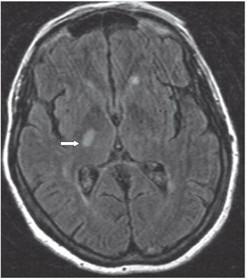
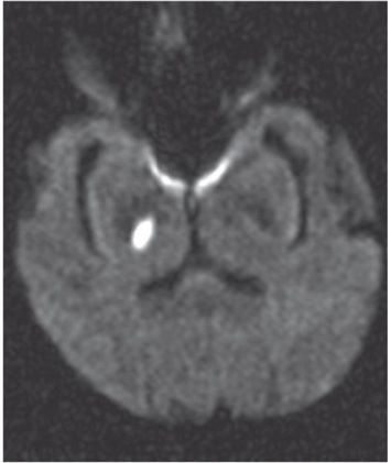
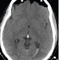
FINDINGS Figure 95-1. Axial NCCT through the basal ganglia. There is a poorly defined hypodensity in the posterior limb of the right internal capsule/anterior thalamus. Figure 95-2. Axial FLAIR MRI through the basal ganglia. There is an ovoid hyperintense lesion (arrow) in the posterior limb of the right internal capsule. Figure 95-3. Corresponding DWI. There is focal restricted diffusion of the internal capsular lesion.
DIFFERENTIAL DIAGNOSIS Enlarged Virchow-Robin spaces, cerebral autosomal dominant arteriopathy with subcortical infarcts and leukoencephalopathy (CADASIL), lacunar infarct, cryptococcus, and neurocysticercosis.
DIAGNOSIS Lacunar infarct (LI) basal ganglia.
DISCUSSION


Stay updated, free articles. Join our Telegram channel

Full access? Get Clinical Tree


