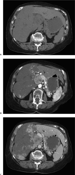
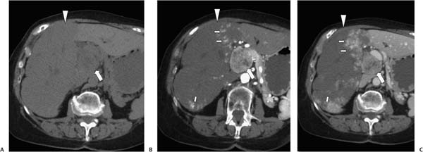
Case 95 A 59-year-old man presents with abdominal pain and weight loss. (A) Noninfused computed tomography (CT) shows a large, hypodense, uncalcified mass (arrowhead) replacing most of the right lobe of the liver. In addition, there is a large, uncalcified mass (arrow) occupying and expanding the porta hepatis. (B) Arterial-phase contrast-enhanced CT shows focal areas of contrast pooling (small arrows) in the periphery of the large hepatic mass (arrowhead). The mass in the porta hepatis (large arrow) occupies the location of the pancreatic head and heterogeneously enhances with pancreatic tissue. The pancreatic body and tail appear atrophied. (C) Venous-phase imaging shows further patchy peripheral enhancement (small arrows) of the large hepatic mass (arrowhead), and diminished enhancement of the tumor in the pancreatic head (large arrow) similar to that in the pancreatic body and tail. • Primary pancreatic carcinoma and cavernous hemangioma of the liver:
 Clinical Presentation
 Imaging Findings
 Differential Diagnosis
![]()
Stay updated, free articles. Join our Telegram channel

Full access? Get Clinical Tree


