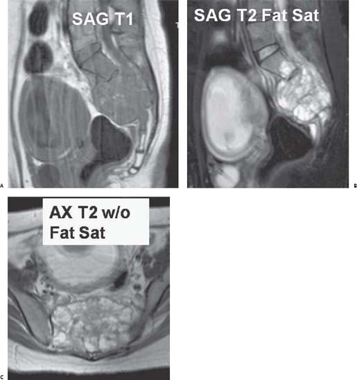
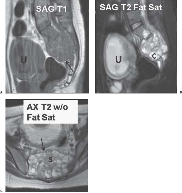
Case 96 The patient is a 19-year-old woman with a mass seen on prior obstetric ultrasonography. (A–C) Magnetic resonance (MR) images show a heterogeneous hyperintense, expansile, mixed cystic (C) and solid (S) mass deforming the sacrum. No internal calcifications or associated soft-tissue mass is grossly evident. A thin, low-signal rim (arrows) encloses the mass. A gravid uterus (U) is present.

 Clinical Presentation
 Imaging Findings

Stay updated, free articles. Join our Telegram channel

Full access? Get Clinical Tree


