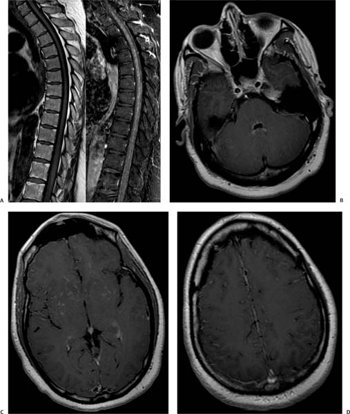
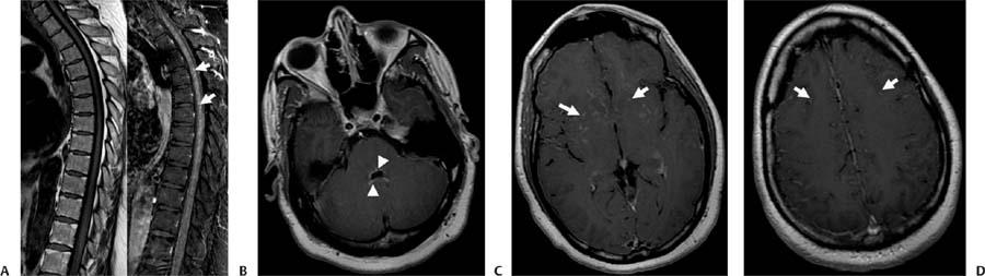
Case 96 A 43-year-old African-American man with a chronic lung condition presenting with the new onset of seizures. (A) Sagittal T1-weighted images (WIs) before and after contrast demonstrate extensive linear leptomeningeal enhancement in the thoracic region (white arrows). (B) Nodular enhancement around the 4th ventricle is demonstrated in the postcontrast axial T1WI (arrowheads). (C) Postcontrast axial T1WI shows the distribution of linear and nodular areas of enhancement in the basal ganglia following the Virchow-Robin spaces (arrows), which is indicative of leptomeningeal involvement. (D) On a postcontrast axial T1WI, leptomeningeal enhancement is also noted in the frontal convexities (arrows). • Leptomeningeal neurosarcoidosis: Sarcoidosis is a diagnosis of exclusion. It is characterized by nodular or linear enhancement of the cortical sulci, perivascular spaces, and cisterns around the base of the brain. • Leptomeningeal carcinomatosis:
Clinical Presentation
Imaging Findings
Differential Diagnosis
![]()
Stay updated, free articles. Join our Telegram channel

Full access? Get Clinical Tree


