 Clinical Presentation
A 45-year-old woman with dyspnea and a heart murmur.
Further Work-up

 Imaging Findings
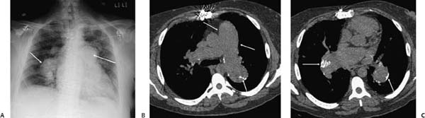
(A) Posteroanterior chest radiograph demonstrates severe enlargement of the main and central pulmonary arteries (arrows). (B) Noncontrast computed tomography (CT; lung windows) at the level of the main pulmonary artery shows significant enlargement of the pulmonary artery. Note the size discrepancy in comparison with the aorta. There is peripheral calcification involving the wall of the left pulmonary artery (arrows
Stay updated, free articles. Join our Telegram channel

Full access? Get Clinical Tree


