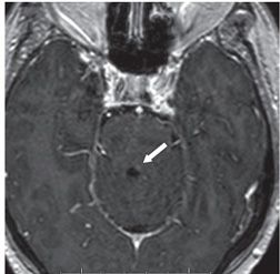
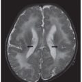
FINDINGS Figure 96-1. Axial T2WI through the medulla. There is hyperintensity within the right medullary olive with minimal swelling (white arrow). Figure 96-2. Axial post-contrast T1WI. The lesion does not show pathologic enhancement to suggest the presence of a metastatic lesion. However, more superiorly, at the level of the contralateral superior cerebellar peduncle, there is a small enhancing focus compatible with metastasis (white arrow).
DIFFERENTIAL DIAGNOSIS Metastatic disease, ischemia, demyelination, hypertrophic olivary degeneration (HOD).
DIAGNOSIS Hypertrophic olivary degeneration (HOD).


Stay updated, free articles. Join our Telegram channel

Full access? Get Clinical Tree


