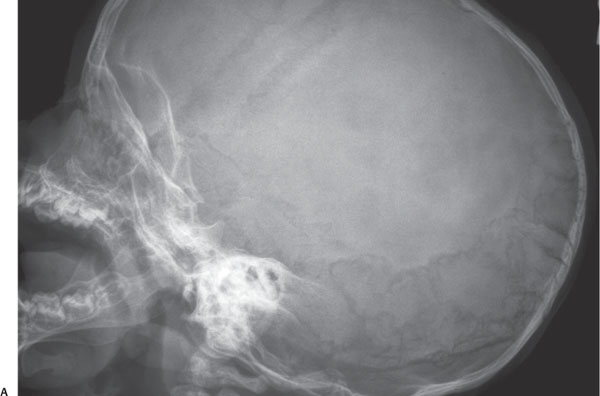
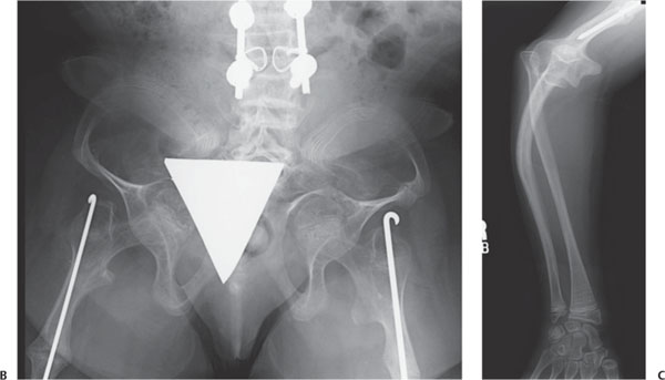
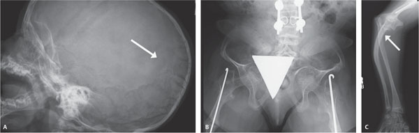
Case 96 A newborn with fractures. Later in life. (A) Lateral view of the skull: there are multiple intrasutural (wormian) bones (arrow). (B) Anteroposterior view of the pelvis: There is severe protrusion of the acetabuli, coxa vara, and an old fracture of the right femur. The femora are thin and contain intramedullary rods. The lumbar vertebral bodies are biconcave and flattened. There are scoliosis rods. (C) Anteroposterior view of the forearm: There is bowing of the ulna (arrow) and a fracture of the distal humerus, which contains an intramedullary rod. There are thin, sclerotic metaphyseal bands parallel to the physis. • Osteogenesis imperfecta (OI): All of the above findings are consistent with OI.
Clinical Presentation
Further Work-up
Imaging Findings
Differential Diagnosis
Stay updated, free articles. Join our Telegram channel

Full access? Get Clinical Tree


